Transplant rejection
 The pathologic mechanisms of transplant rejection are complex, and they can be hard to get a grip on. Here are some questions that may help you understand these processes a little better. (more…)
 The pathologic mechanisms of transplant rejection are complex, and they can be hard to get a grip on. Here are some questions that may help you understand these processes a little better. (more…)

Factor V Leiden is a genetic disorder in which patients have an increased tendency to form thromboses, or blood clots. (more…)
Here’s an example of a common question students have in the beginning of a medical school or dental school pathology course.
Unfortunately, students often feel like they “should” know the answers to certain questions – so they don’t ask. Don’t fall into this trap! You never need to feel embarrassed about asking a question; everyone has things they don’t know – even professors. That’s why you’re taking the class – to learn!
On to the question.
Q. What is the main difference between a neutrophil and a monocyte? This is what I understand:
PMNs:
fight bacteria and fungi (but they are different than NK cells–right?)
act as antigen presenting cells
phagocytic
are generally the first to arrive; part of the acute inflammatory response
Monocytes:
act as antigen presenting cells
can secrete cytokines and attract inflammatory cells like fibroblasts, etc.
phagocytic
bigger role in chronic inflammation
A. Broadly, the similarities are: neutrophils and monocytes are both phagocytes, and they both work to fight infections. But moncytes can turn into macrophages (when they get into tissues), which are very good at eating things, as well as presenting antigens. Neutrophils eat, but don’t present, antigens. One of the big differences, too, you already mentioned: neutrophils are the first to come in during an inflammatory process. Lymphocytes come next, then monocytes/macrophages come in to mop up the mess.
One note: neutrophils are phagocytes, but not antigen presenting cells. Another note: You are right, neutrophils are different than NK cells. NK (natural killer) cells are specialized lymphocytes which have functions different than those of neutrophils and monocytes.
Also: neutrophils look different than monocytes/macrophages. Neutrophils have a “busy” nucleus (that’s why they are called “polymorphonuclear” leukocytes), with several lobes. You can see one at 2 o’clock in the above photo. They also have granules, both primary (azurophilic) and secondary (fawn-colored). Monocytes have a horseshoe-shaped nucleus, with dishwater-gray cytoplasm and a few tiny granules. See the lower left corner in the above photo.
There are four types of thyroid carcinoma: papillary, follicular, medullary, and anaplastic carcinoma. (more…)
Papillary thyroid carcinoma has a number of unique morphologic features. I mentioned psammoma bodies a few days ago. (more…)
Just as there are many different types of myeloid cells (neutrophils, red cells, monocytes, eosinophils, basophils), there are many different types of acute myeloid leukemia (AML). Two types of AML are composed almost entirely of cells of the monocytic series: acute monoblastic leukemia and acute monocytic leukemia. In both of these types of AML, at least 80% of the leukemic cells are from the monocytic series (monoblasts, promonocytes, and monocytes). In acute monoblastic leukemia, most of these cells are monoblasts, and in acute monocytic leukemia, most of these cells are promonocytes. Promonocytes have a very characteristic appearance, as shown above. They have nuclei that show a delicate folding pattern, almost like a piece of tissue paper that has been crumpled a bit. If you had a case of acute leukemia and most of the cells looked like this, you would think about acute monocytic leukemia – and you’d get an NSE to prove it.

Some cases of acute leukemia are composed entirely of undifferentiated-appearing blasts. These cases are difficult (or impossible) to diagnose morphologically (under the microscope) – you really need special tests like immunophenotyping in order to make a definitive diagnosis. (more…)
 One of the characteristic features of papillary thyroid carcinoma is the presence of psammoma bodies. These are calcifications with an unusual (and pretty) lamellar pattern. (more…)
When you’re faced with an acute leukemia composed entirely of blasts, one way to figure out the identity of those blasts is to use cytochemical stains. (more…)
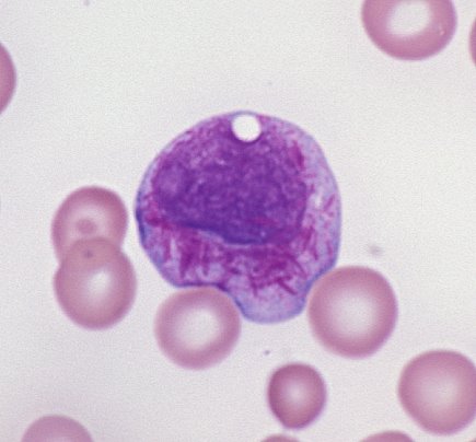
Some types of acute leukemia are composed of only blasts (no differentiating neutrophils, no monocytic precursors, just a sea of blasts). In those cases, look for Auer rods. A blast with an Auer rod can only be a myeloblast! It cannot be a lymphoblast, or a monoblast, or any other kind of blast. So if you see blasts with Auer rods, you know it is some type of acute myeloid leukemia. Remember, though, that the converse is not true: just because you don’t see Auer rods, that does not mean that the blast is not a myeloblast. Some myeloblasts have Auer rods, and some don’t. So if you see Auer rods, it is an AML. If you don’t, it still could be an AML.
Recent Comments