What exactly does “storiform” mean?

Do you know what a “storiform” pattern is? Yeah, neither did I when I was a medical student. However, that term did get thrown around in pathology lectures a lot, without any description or definition. There are lots of terms like this – so I’m gonna just go ahead and create a new category called:

Words Pathologists Use In Lecture As If You Know What They Mean.

I think it’s important to pause and define these terms, because otherwise this is what happens: given the sheer volume of stuff you’re supposed to learn, and the minimal amount of time you have to accomplish this task, you’re not going to look up every word you have a nagging doubt about. You’re going to infer the meaning from whatever was said in lecture, and wind up with a fuzzy and probably incorrect definition. And then someone will ask you about it on rounds, and it will be frustrating.

SO. We’ll start with “storiform” today – and I’ll keep adding posts about terms in this category as I run across them. Please email me if you find a word like this! Then we’ll wind up with a nice glossary of these formerly-unexplained terms, and you’ll look like the star you are when one of these terms comes up on rounds.

First, a little Latin

Storiform comes from the Latin storea (woven mat) and formis (form, or pattern) – so technically, storiform means “having the pattern of a woven mat.” When we use “storiform” in pathology, though, it has a more specific meaning. It refers to a tumor pattern consisting of spindle cells in a pinwheel-shaped arrangement (radiating out from a central core).

What does it look like?

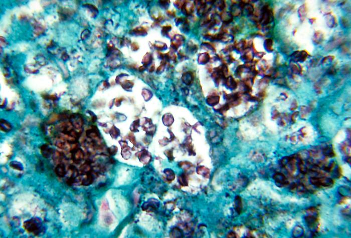
Sooo…what does a pinwheel-shaped arrangement of spindle cells look like under the microscope? Let’s try looking at an un-marked slide first, just to see if you can find pinwheels on your own. Here’s an image (below, right) of a skin lesion called dermatofibrosarcoma protuberans (DFSP), which is known for its storiform pattern. Take a look and see if you can find areas where the tumor cells are arranged in a pinwheel-shaped fashion. Then scroll down to see a labeled image.

Ready to see the labeled image? Okay, scroll down….

.

.

.

.

.

.

.

.

Keep going…

.

.

.

Here’s the same image, faded out a bit so you can see the pinwheel-shaped areas outlined in black:

Okay, they’re not perfectly symmetrical pinwheels, but they do look pinwheel-ish, with tumor cells radiating out from a central core region.

One last thing, now that we’ve got the pinwheels down…if you go back and look at the unmarked image again, you might be able to imagine that the cells are arranged like a woven mat, like the Latin term suggests.

I hope you feel comfortable with “storiform” now. That’s one less undefined term!

How do gallstones form?

There are two types of gallstones: cholesterol stones and pigment stones. If you didn’t know anything about gallstones, you’d guess (rightly so) that cholesterol stones are made up of cholesterol. And you’d also probably guess that you get cholesterol stones when there’s too much cholesterol around. But how, exactly? And pigment stones – what are those made of? Pigment?

Turns out there are very good explanations for all of these questions. Let’s take a look.

Cholesterol gallstones

Cholesterol gallstones contain – not surprisingly – cholesterol. And they arise when there’s more cholesterol around than the gallbladder can handle. But what does this actually mean?

A tiny bit of basic science here. Under normal conditions, cholesterol is soluble in bile because it binds to bile salts (which are water-soluble) and lecithins (which are water-insoluble). These guys both act like detergents, and cholesterol is dispersed within the bile, and everything’s cool.

But what happens if there’s too much cholesterol around? If the concentration of cholesterol exceeds the solubilizing capacity of bile, then cholesterol will nucleate into solid cholesterol crystals, which can over time get big enough to form stones.

Pigment gallstones

These stones are made of unconjugated bilirubin (mixed with calcium salts). They’re called pigment gallstones because they’re dark brown to black in color (compared to cholesterol stones, which are usually pale yellowish-greenish in color).

The two main conditions in which you see pigment stones are chronic hemolytic anemia and infection of the biliary tract. Why would these conditions lead to an accumulation of unconjugated bilirubin in the bile? In order to answer that, let’s quickly review bilirubin metabolism in the bile itself.

Normally, the liver conjugates bilirubin and dumps it into the bile. So the bile contains just conjugated bilirubin, then, right? Wrong! About 1% of the bilirubin in bile undergoes deconjugation while it’s still in the biliary tree (betcha didn’t know that!). Bile is then dumped into the gut, where bacterial-glucuronidases convert most of the remaining conjugated bilirubin into its unconjugated form.

Back to the causes of pigmented stones. If you have an infection in the biliary tree, and the infectious agent makes glucuronidase, then you’ll end up deconjugating more bilirubin than normal…and over time, that unconjugated bilirubin can accumulate and form stones.

The other main cause of pigmented stones is chronic hemolysis. If you’re busting open lots of red cells, all that heme gets transformed into bilirubin, which the liver conjugates and dumps into the bile. So the bile contains a lot more bilirubin than usual. Most of that bilirubin remains conjugated – but around 1% is turned into unconjugated bilirubin right there in the biliary tree. If you’re making a lot more bilirubin than normal, that 1% is significant – and over time, that excess of unconjugated bilirubin can be enough to lead to pigment stones.

Name that organism!

Can you identify this organism?

Wow, I got lots of good feedback on the last post I did with a “name that bug” theme – so I’m going to do more! I like learning this way – especially when there’s no one around to judge you. Unknown conferences during pathology residency could be pretty brutal…but we’re all friends here – so if you don’t get the answer right, it’s not a problem – it’s just an opportunity to learn something new. How nice.

Okay. Start by taking a look at this image – maybe you’ll know right away what it is, and maybe you won’t. If you want more hints, just scroll down a little and keep reading.  The answer is at the bottom – so don’t scroll way down until you’re ready.

How do you get it?

Our mystery organism makes its home in soil. It particularly likes damp soil that’s rich with decomposing stuff (like wood and leaves). In the US, it’s seen mostly in central eastern and southeastern states (if you draw a line from the western border of Minnesota down to the eastern border of Texas, this organism doesn’t really like to live west of that line). It’s seen in Canada too.

What are the symptoms?

This organism typically just affects the lungs – but some patients do develop disseminated disease. Very rarely, the organism directly infects the skin, and just causes problems there. Most patients present with abrupt-onset productive cough, fever, chills, and chest pain. It may resolve on its own, or hang around and become chronic.

What does it look like?

The main thing you see under the microscope with this organism is suppurative (pus-filled) granulomas. Macrophages aren’t so great at killing this organism – so neutrophils come to the rescue (they’re the main cellular constituent of pus).

The organism itself is a round, with a thick (some say “double-contoured” but our photo doesn’t show that) cell wall, and – here’s the kicker – broad-based budding. In a histologic section, like this one (which is stained with PAS, by the way), you can see nice big nuclei in each round organism.

The cells in green circles are neutrophils (you can tell by their “busy” nuclei that look like Mickey Mouse ears); the yellow arrow points to the thick cell wall, the red arrows point to the nuclei, and the red oval points out the broad-based budding between two organisms.

Okay. Ready for the answer? Scroll down…


Keep going!


This organism is Blastomyces dermatitidis! Normally, I’d link the image itself – but in this case, I didn’t want you to accidentally see the answer…so here’s the link.

Here’s a recap of the main things to remember about Blastomyces and blastomycosis:

  • Lives in decomposing soil
  • Ohio and Mississippi river valleys
  • Usually just causes pneumonia, but can become disseminated
  • Rarely, localized to skin (which is probably why it got the name dermatitidis)
  • Suppurative granulomas
  • Large, round, thick-walled organisms with broad-based budding

Finally, as an aside, I remember when we were learning this in med school, we put a capital “B” by the organism, because it’s Blastomyces (obviously), and:

  • It’s pretty big
  • It shows broad-based budding
  • When it buds, it kind of looks like a capital B if you use your imagination.


Name that organism!

Can you identify this organism?

Start by taking a look at the image – and if you need more hints (it’s okay if you do!), keep reading. The answer is at the bottom – so don’t scroll down until you’re ready.

How do you get it?

Let’s start by talking about the places you’re most likely to encounter our mystery organism. This little guy likes to hang out in contaminated dust or soil. The most common means of contamination is through bird or bat droppings (yuck). The organism is acquired by inhalation – so spelunkers (who might breathe in soil with bat poopy) and construction workers (who might breathe in dust with bird poopy) are at increased risk.

Endemic areas in the US include the Ohio and Mississippi rivers. Outside the US, this organism is endemic in the Caribbean – but it’s also found in a bunch of other places (Mexico, Central and South America, eastern/southern Europe, Africa, eastern Asia, and Australia). I hate these long lists, btw. I mean, you might as well memorize the places this organism is NOT found. In these situations, it’s best to just memorize the endemic places first, and then if there’s room in your head later, you can stick in the other places.

What are the symptoms?

This organism can produce several different clinical patterns of disease, including:

  • Focal, self-limiting lung lesions with mild or no symptoms
  • Chronic, progressive lung disease with cough, fever, and night sweats
  • Disseminated disease

Although disease can occur in immunocompetent patients, it’s more common (and more severe) in patients who are immunocompromised.

What does it look like?

The main histologic abnormality this organism causes is granulomas. Non-immunocompromised patients get caseating granulomas which tend to undergo calcification after a while. Immunocompromised patients can’t really form nice granulomas, especially if they have diminished T cell function (because you need working T cells to make granulomas). So in these patients, you’ll just see clumps of organism-containing phagocytic cells here and there.

The organism itself is a cute little guy – emphasis on little. In fact, it’s so tiny that you really have to go on high power to see it. It’s usually seen in macrophages (weird place to hang out, unless you have a death wish). It’s spherical in shape, and its walls are thin.

Here’s a labeled image so you can see what’s going on. The red circles all show macrophages stuffed with varying numbers of organisms and/or cut in varying planes of section. What you’re really seeing is the cytoplasm of these macrophages (it’s often hard to see macrophage nuclei, especially when they’re so stuffed). There are TONS of organisms, but I’ll just point out one really good one (red arrow) to sear into your brain for future reference.

Okay. Ready for the answer? Scroll down…


Keep going!


This organism is Histoplasma capsulatum! Normally, I’d link the image itself – but in this case, I didn’t want you to accidentally see the answer…so here’s the link. The CDC has a lot of great images, by the way, if you’re ever looking for one for a presentation.

Here’s a recap of the main things to remember about Histoplasma and histoplasmosis:

  • Inhalation of dirt with bird/bat poopy
  • Ohio and Mississippi rivers, Caribbean
  • Asymptomatic, or chronic lung disease, or disseminated disease
  • Worse in immunocompromised patients
  • Cute, tiny, round organisms in macrophages


What does megaloblastic mean?

Here are a few great questions about megaloblastic anemia I received by email.

Megaloblastic vs. macrocytic

Q. Do I have to say “megaloblastic macrocytic” anemia? Aren’t megaloblastic and macrocytic the same thing?

A. Macrocytic refers to the size of the mature red cells in the blood. It means that the red cells are big. Normal is 80-100 femtoliters. If the red cells are over 100, they’re macrocytic; if they’re under 80, they’re microcytic.

Megaloblastic refers to the weird morphologic changes (immature nucleus, mature cytoplasm, large overall size) you see in red cell precursors (and, to some extent in neutrophil precursors), in patients who are B12 deficient. So the term is really referring to the cells in the bone marrow, not mature, circulating red cells. However, you can also see changes in the blood that indicate megaloblastic anemia, the most common of which is hypersegmented neutrophils (like the one above).

So the terms are not equivalent.

That being said, you don’t need to say both terms if you have a megaloblastic anemia, because all megaloblastic anemias are also macrocytic. You just say “megaloblastic anemia.”

Conversely, if you just say “macrocytic anemia,” that doesn’t say anything about whether there are megloblastic changes present or not! It just says: there’s an anemia, and the red cells are big.

Non-megaloblastic anemia

Q. What really is non-megaloblastic anemia? Because my lectures have mentioned it but I’m not sure what it really is.

A. Non-megaloblastic anemia just means an anemia without megaloblastic changes – and technically, that encompasses every single anemia except megaloblastic anemia! But really, when people say non-megaloblastic anemia, they’re usually referring to a macrocytic anemia (one in which the red cells are large, over 100 femtoliters) without megaloblastic changes (funny looking red cells). This type of anemia can be seen in liver failure and in myelodysplasia.

Pernicious anemia and megaloblastic anemia

Q. I don’t understand the difference between pernicious anemia and megaloblastic anemia. Pernicious anemia is just a deficiency in intrinsic factor that helps with absorption of B12…so patients have low B12 levels. But how is that different from megaloblastic anemia?

A. The best way to think about these two terms is: pernicious anemia is one cause of megaloblastic anemia.

Megaloblastic anemia is a type of anemia in which you get weird morphologic changes (megaloblasts, hypersegmented neutrophils, oval macrocytes) due to a lack of B12 and/or folate. There are lots of things that can cause a lack of B12 and/or folate…so when you see a case of megaloblastic anemia, you have to investigate to find out what the cause is.

Pernicious anemia (in which patients can’t absorb B12 due to a lack of intrinsic factor) is one cause. Another cause is folate-depleting drugs (like chemotherapy drugs); another is dietary deficiency.

It’s kind of confusing because they put the term “anemia” in pernicious anemia – so it makes it sound like pernicious anemia is a category in and of itself. It’s not – it just falls under the heading of megaloblastic anemia.