Can you identify this organism?
Start by taking a look at the image – and if you need more hints (it’s okay if you do!), keep reading. The answer is at the bottom – so don’t scroll down until you’re ready.
How do you get it?
Let’s start by talking about the places you’re most likely to encounter our mystery organism. This little guy likes to hang out in contaminated dust or soil. The most common means of contamination is through bird or bat droppings (yuck). The organism is acquired by inhalation – so spelunkers (who might breathe in soil with bat poopy) and construction workers (who might breathe in dust with bird poopy) are at increased risk.
Endemic areas in the US include the Ohio and Mississippi rivers. Outside the US, this organism is endemic in the Caribbean – but it’s also found in a bunch of other places (Mexico, Central and South America, eastern/southern Europe, Africa, eastern Asia, and Australia). I hate these long lists, btw. I mean, you might as well memorize the places this organism is NOT found. In these situations, it’s best to just memorize the endemic places first, and then if there’s room in your head later, you can stick in the other places.
What are the symptoms?
This organism can produce several different clinical patterns of disease, including:
- Focal, self-limiting lung lesions with mild or no symptoms
- Chronic, progressive lung disease with cough, fever, and night sweats
- Disseminated disease
Although disease can occur in immunocompetent patients, it’s more common (and more severe) in patients who are immunocompromised.
What does it look like?
The main histologic abnormality this organism causes is granulomas. Non-immunocompromised patients get caseating granulomas which tend to undergo calcification after a while. Immunocompromised patients can’t really form nice granulomas, especially if they have diminished T cell function (because you need working T cells to make granulomas). So in these patients, you’ll just see clumps of organism-containing phagocytic cells here and there.
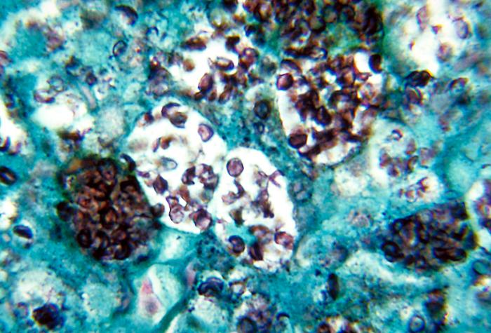
The organism itself is a cute little guy – emphasis on little. In fact, it’s so tiny that you really have to go on high power to see it. It’s usually seen in macrophages (weird place to hang out, unless you have a death wish). It’s spherical in shape, and its walls are thin.
Here’s a labeled image so you can see what’s going on. The red circles all show macrophages stuffed with varying numbers of organisms and/or cut in varying planes of section. What you’re really seeing is the cytoplasm of these macrophages (it’s often hard to see macrophage nuclei, especially when they’re so stuffed). There are TONS of organisms, but I’ll just point out one really good one (red arrow) to sear into your brain for future reference.

Okay. Ready for the answer? Scroll down…
Keep going!
This organism is Histoplasma capsulatum! Normally, I’d link the image itself – but in this case, I didn’t want you to accidentally see the answer…so here’s the link. The CDC has a lot of great images, by the way, if you’re ever looking for one for a presentation.
Here’s a recap of the main things to remember about Histoplasma and histoplasmosis:
- Inhalation of dirt with bird/bat poopy
- Ohio and Mississippi rivers, Caribbean
- Asymptomatic, or chronic lung disease, or disseminated disease
- Worse in immunocompromised patients
- Cute, tiny, round organisms in macrophages


Wow. This is such a cool way to learn organisms. Thanks.
This format is awesome!!!!
I am loving these readings. Well written and very organized.
Really enjoyed the info, just enough to keep you being excited for the next one.
The information really help me a lot. Will you please discuss some things about Body Fluids especially the microscopic parts.
Thank you,
Mercedes