Blood cookies!
I’ve been really busy teaching this fall, so I haven’t been posting nearly as much as I’d like. I will be back to normal (ha) soon – but until then, I thought I’d share what we did in class yesterday. We’ve been learning about hematopathology (my favorite) – so I made cookies depicting some of the diseases we covered.


It’s super geeky, but I’m okay with that. It’s really fun to combine path knowledge with something that’s actually creative and pretty. And it’s sort of educational for my class…at the very least, they get a well-deserved break from the HOURS of lecture they have to sit through. Here are the end results (with a few high-yield things about each cell).

Sickle cells
Sickle cells are seen, of course, in sickle cell anemia. They’re abnormally shaped because when sickle hemoglobin deoxygenates, it polymerizes, contorting the red cell into a sickle shape.

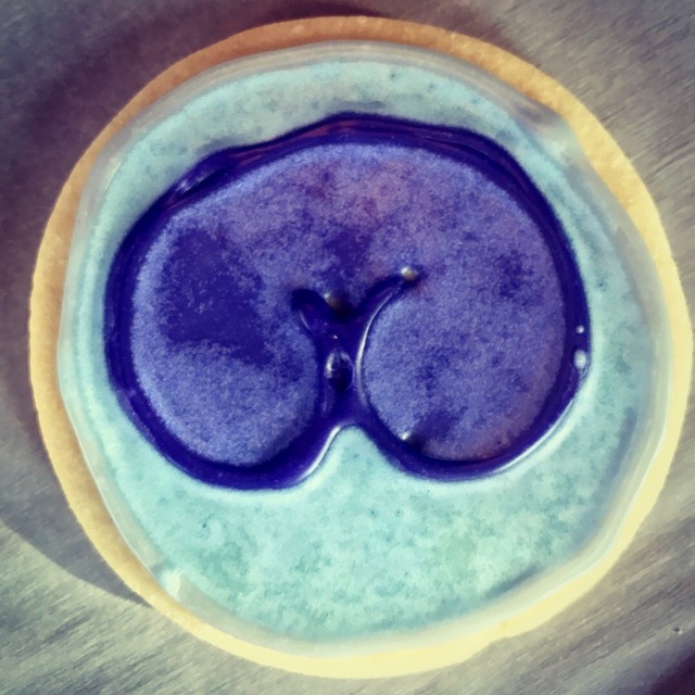
Reed-Sternberg cell
Reed-Sternberg cells are the malignant cells in Hodgkin lymphoma. They’re gigantic, and typically they have two nuclei with prominent nucleoli, giving the cell an “owl’s-eye” appearance.

Neutrophil with Döhle body
In some cases of bacterial infection, neutrophils develop little blue cytoplasmic inclusions, called Döhle bodies, which are chunks of revved-up rough endoplasmic reticulum.
Butt cell
No, I didn’t make this up! Follicular lymphoma is made up, in part, of small cleaved cells – and when these get out into the blood, their nuclei totally look like butts. Adorable.

Faggot cell
Faggot cells contain TONS of Auer rods (faggot means bundle of sticks). They’re pathognomonic of acute promyelocytic leukemia, which has a t(15;17) that you should stick in your head somewhere.

Blast with Auer rod
Auer rods are only seen in malignant myeloblasts. So if you see one, you know you’re dealing with acute myeloid leukemia. Not all AMLs have Auer rods, though – so the absence of Auer rods doesn’t rule out AML.

Platelets
These could be normal platelets…but since we’re talking about diseases, let’s say they’re platelets from essential thrombocythemia, which is one of the four chronic myeloproliferative disorders (it’s the one in which the blood has an extremely high platelet count).


Recent Comments