Does pyknosis occur in necrosis or apoptosis?

Q: Would a pyknotic cell be a form of necrosis or apoptosis? Or am I totally off base here?

A. No you’re totally not off base – that’s a really good question!!

We typically use the word “pyknosis” to mean one of the three nuclear patterns seen in necrotic cells…but pyknosis can also occur in apoptosis! I’ll explain a bit more.

When cells undergo necrosis, they show a lot of different morphologic abnormalities. Overall, necrotic cells appear enlarged and more eosinophilic, and their nuclei look abnormal due to breakdown of DNA. There are three specific patterns of nuclear change in necrosis, which are:

  • Pyknosis (the nucleus shrinks and becomes dark blue/black)
  • Karyorrhexis (the nucleus breaks apart, or fragments, like a cookie crumbling into bits)
  • Karyolysis (the nucleus just fades away)

Here’s a nice diagram I found (I modified it a bit from the original) showing these changes:
When cells undergo apoptosis, they also show a lot of different morphologic features. Overall, apoptotic cells appear shrunken, with really dense, dark, eosinophilic cytoplasm, and the chromatin in the nucleus aggregates into a dense mass which can fragment. Here’s a photo from Robbins showing an apoptotic cell:

The word “pyknosis” isn’t typically used when describing an apoptotic cell – but Robbins does say that pyknosis can be a feature of apoptotic cells, so there you go.

So…the bottom line is that pyknosis is a nuclear change in which the nucleus shrinks and becomes dark blue/black. Typically, we associate the word “pyknosis” with necrotic cells – but apoptotic cells can show pyknosis too.

Name that organism!

Can you identify this organism?

Wow, I got lots of good feedback on the last post I did with a “name that bug” theme – so I’m going to do more! I like learning this way – especially when there’s no one around to judge you. Unknown conferences during pathology residency could be pretty brutal…but we’re all friends here – so if you don’t get the answer right, it’s not a problem – it’s just an opportunity to learn something new. How nice.

Okay. Start by taking a look at this image – maybe you’ll know right away what it is, and maybe you won’t. If you want more hints, just scroll down a little and keep reading.  The answer is at the bottom – so don’t scroll way down until you’re ready.

How do you get it?

Our mystery organism makes its home in soil. It particularly likes damp soil that’s rich with decomposing stuff (like wood and leaves). In the US, it’s seen mostly in central eastern and southeastern states (if you draw a line from the western border of Minnesota down to the eastern border of Texas, this organism doesn’t really like to live west of that line). It’s seen in Canada too.

What are the symptoms?

This organism typically just affects the lungs – but some patients do develop disseminated disease. Very rarely, the organism directly infects the skin, and just causes problems there. Most patients present with abrupt-onset productive cough, fever, chills, and chest pain. It may resolve on its own, or hang around and become chronic.

What does it look like?

The main thing you see under the microscope with this organism is suppurative (pus-filled) granulomas. Macrophages aren’t so great at killing this organism – so neutrophils come to the rescue (they’re the main cellular constituent of pus).

The organism itself is a round, with a thick (some say “double-contoured” but our photo doesn’t show that) cell wall, and – here’s the kicker – broad-based budding. In a histologic section, like this one (which is stained with PAS, by the way), you can see nice big nuclei in each round organism.

The cells in green circles are neutrophils (you can tell by their “busy” nuclei that look like Mickey Mouse ears); the yellow arrow points to the thick cell wall, the red arrows point to the nuclei, and the red oval points out the broad-based budding between two organisms.

Okay. Ready for the answer? Scroll down…


Keep going!


This organism is Blastomyces dermatitidis! Normally, I’d link the image itself – but in this case, I didn’t want you to accidentally see the answer…so here’s the link.

Here’s a recap of the main things to remember about Blastomyces and blastomycosis:

  • Lives in decomposing soil
  • Ohio and Mississippi river valleys
  • Usually just causes pneumonia, but can become disseminated
  • Rarely, localized to skin (which is probably why it got the name dermatitidis)
  • Suppurative granulomas
  • Large, round, thick-walled organisms with broad-based budding

Finally, as an aside, I remember when we were learning this in med school, we put a capital “B” by the organism, because it’s Blastomyces (obviously), and:

  • It’s pretty big
  • It shows broad-based budding
  • When it buds, it kind of looks like a capital B if you use your imagination.


Name that organism!

Can you identify this organism?

Start by taking a look at the image – and if you need more hints (it’s okay if you do!), keep reading. The answer is at the bottom – so don’t scroll down until you’re ready.

How do you get it?

Let’s start by talking about the places you’re most likely to encounter our mystery organism. This little guy likes to hang out in contaminated dust or soil. The most common means of contamination is through bird or bat droppings (yuck). The organism is acquired by inhalation – so spelunkers (who might breathe in soil with bat poopy) and construction workers (who might breathe in dust with bird poopy) are at increased risk.

Endemic areas in the US include the Ohio and Mississippi rivers. Outside the US, this organism is endemic in the Caribbean – but it’s also found in a bunch of other places (Mexico, Central and South America, eastern/southern Europe, Africa, eastern Asia, and Australia). I hate these long lists, btw. I mean, you might as well memorize the places this organism is NOT found. In these situations, it’s best to just memorize the endemic places first, and then if there’s room in your head later, you can stick in the other places.

What are the symptoms?

This organism can produce several different clinical patterns of disease, including:

  • Focal, self-limiting lung lesions with mild or no symptoms
  • Chronic, progressive lung disease with cough, fever, and night sweats
  • Disseminated disease

Although disease can occur in immunocompetent patients, it’s more common (and more severe) in patients who are immunocompromised.

What does it look like?

The main histologic abnormality this organism causes is granulomas. Non-immunocompromised patients get caseating granulomas which tend to undergo calcification after a while. Immunocompromised patients can’t really form nice granulomas, especially if they have diminished T cell function (because you need working T cells to make granulomas). So in these patients, you’ll just see clumps of organism-containing phagocytic cells here and there.

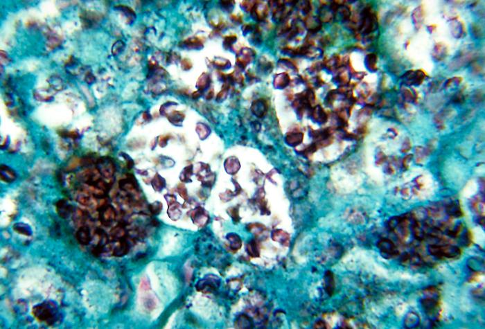
The organism itself is a cute little guy – emphasis on little. In fact, it’s so tiny that you really have to go on high power to see it. It’s usually seen in macrophages (weird place to hang out, unless you have a death wish). It’s spherical in shape, and its walls are thin.

Here’s a labeled image so you can see what’s going on. The red circles all show macrophages stuffed with varying numbers of organisms and/or cut in varying planes of section. What you’re really seeing is the cytoplasm of these macrophages (it’s often hard to see macrophage nuclei, especially when they’re so stuffed). There are TONS of organisms, but I’ll just point out one really good one (red arrow) to sear into your brain for future reference.

Okay. Ready for the answer? Scroll down…


Keep going!


This organism is Histoplasma capsulatum! Normally, I’d link the image itself – but in this case, I didn’t want you to accidentally see the answer…so here’s the link. The CDC has a lot of great images, by the way, if you’re ever looking for one for a presentation.

Here’s a recap of the main things to remember about Histoplasma and histoplasmosis:

  • Inhalation of dirt with bird/bat poopy
  • Ohio and Mississippi rivers, Caribbean
  • Asymptomatic, or chronic lung disease, or disseminated disease
  • Worse in immunocompromised patients
  • Cute, tiny, round organisms in macrophages


Fuscus, February.

Before I get to fuscus, I have to say that I have HAD it with this month. Normally I start complaining about February around mid-January, and continue on every day until people start avoiding me. This year, I thought I could rise above it. I tried thinking positively. I tried reading Carl Jung and Viktor Frankl to address my existential crisis. Last night I was up late listening to Jordan Peterson, and I thought I was doing pretty well.

But this morning, after coming out to a car completely encased in ice (not exaggerating), after seeing the sky the same color as the ground (greyish white) YET AGAIN, and after some other crap that I won’t go on about, I’m done. February just sucks. It’s not me, it’s you, February.

So where did February come from?

Since I sat down intending to write about the very nice Latin word fuscus, I thought maybe I’d look up the origin of the word February and see if there was some Latin that could help make me feel better. And there was! So I’m sharing what I found in the hopes that it may help others who have February issues.

February is usually said to be derived from the Latin februum, meaning anything that purifies or consecrates. This doesn’t fit my view of this month, but whatever. According to a prominent Roman grammarian (what a cool job, seriously) named Censorinus, who wrote De Die Natali (The Birthday Book) in 238 AD, there were tons of februamenta (rites of purification) going on in Rome during this time of the calendar year. Part of the purification was agricultural (clearing away dead stuff and getting the fields ready for planting). But it wasn’t just fields – houses, material items, and even the Romans themselves were februa (purified) in different ways, using different rites.

Wait, why all the purification?

In Roman tradition, February was originally the last month of the year, and, according to Ovid, it was “consecrated to the shades of the dead.” So along with acts of propitiation (appeasement, I had to look it up too), there were acts of purification to protect the living from evil spirits and also to banish the spirits at the end of the year so that the new year could begin in purity.

The image above is a drawing of the month of February from a fourth century calendar. The caption describes how to placate the ghosts that roam the earth in February. Okay – keeping angry ghosts from haunting the living – now we’re getting somewhere.

There must be something better, though.

Yes, there is. Some sources suggest that February actually comes from feber, a word which the Romans used to describe lamentation. YES! That’s it. I KNEW there had to be something in the name of this miserable month that reflected its true nature. Okay, the Romans were lamenting their deceased. But still. It fits. For the rest of the month, I’m going to say Lamentation instead of February and see if anyone notices.

Okay, back to fuscus.

For those of you brave enough to read this far, here’s a happy thing to offset all the misery. As you may know, there are a bunch of pigments you can see sometimes in tissues. One of them is called lipofuscin. It’s known as the wear-and-tear pigment, because it accumulates with age. Lipofuscin is composed of a bunch of lipids and proteins and has a yellow-brown appearance. It has no clinical significance – but you need to know it exists so you don’t confuse it with something else yellow-brown, like hemosiderin.

Here’s the good part. The first half of lipofuscin makes sense, since it’s composed partly of lipids. The second half is derived from the Latin word fuscus, which means dingy, brown, or dark – great Latin word choice, since lipofuscin has a dingy, brown appearance. So that’s why “obfuscate” means “to make unclear or obscure.” And it may be where we got the word “dusk” (from Middle English dosk, which came from Old English dox, which probably came from our Latin hero fuscus).

This is the weird kind of thing that makes me happy. I have no idea why finding the Latin connection between two seemingly unrelated words should make me happy in such disproportionate measure – or happy at all – but it does.

In this month of Lamentation, I’ll take happiness where I can get it.

What does “differentiation” mean?


Q. I have Googled and YouTubed this thing to death, and I still can’t grasp the meaning of “differentiation.” It seems the opposite of its definition. To “differentiate” means to recognize what makes something different. But according to your post on tumor differentiation, well-differentiated tumors resemble (don’t look different from) their tissue of origin. I would think if something is well-differentiated, it would look very different from the thing it’s being compared to. Why is the use here opposite of its meaning?

A. I totally get where you’re coming from. It’s REALLY frustrating in pathology when things are described in terms that don’t seem to make sense. You are not alone in questioning the use of this term!

The problem is that the word in question – differentiation – has a specific meaning in the real world. You’re exactly right in your definition: to differentiate between two things means to recognize what’s different or unique.

So you’d think that “differentiation” in the pathology world would mean the same thing: the recognition of things that are unique, different, or not the same. By logical reasoning, then, a “differentiated” tumor would be one that looked different from its cell of origin. And you’d think a “well-differentiated” tumor would be one that looked very different from its cell of origin.

Unfortunately, “differentiation” doesn’t have the same definition in the pathology world. So we have to put aside our logic and knowledge of vocabulary for a moment, irritating as that may be, and learn a new definition for this word.

The definition of “differentiation” in pathology-speak.

When we’re talking about tumors, the definition of “differentiation” is simply this: the degree to which tumor cells resemble their cell of origin. A well-differentiated tumor is one in which the tumor cells look very much like their cell of origin. A poorly-differentiated tumor (like the poorly-differentiated squamous cell carcinoma shown above) is one in which the tumor cells barely resemble their cell of origin.

That’s it. Yes, it’s an annoying word choice, because it is used here in a way that seems counterintuitive. But maybe it’s not as far off as it seems.

Maybe this will help.

I think about it (okay, rationalize it) this way. When cells are really immature, they don’t have a lot of features that make them look different from other cells. Myeloblasts don’t look very different than lymphoblasts, for example. So we could say that these immature cells are undifferentiated; it’s hard to tell what kind of cell they really are, and hard to tell them apart from other cells.

The same thing is true of the cells in poorly-differentiated tumors! The cells show practically no features that give away their identity; it’s hard to even tell what kind of cells they are. They are, in effect, undifferentiated.

If you think about “differentiation” this way (undifferentiated cells lack identifying features; it’s hard to tell what kind of cell they are), then the concept of tumor differentiation is a little easier to swallow. A little.

Tumor invasion and metastasis: are they the same thing?

Here are a couple great questions from one of my lovely students regarding invasiveness and metastasis.

Q. I have a quick question on today’s lecture. There is a slide near the end that has a picture of non-invasive carcinoma. For a tumor to be malignant, should it not be invasive? (more…)